Fungal infections of the skin and hair are often caused by dermatophytes such as Trichophyton or Microsporum. They can be spread between people (particularly children) or transmitted from animals or soil. In very rare cases they can become invasive in patients who are immunodeficient (Wang et al, 2020). However, even superficial skin infections can cause distress and social stigma.
- Read more at GAFFI: 138 million school age children in Africa have fungal scalp infection, affecting education and leading to social stigmas
When sending a specimen for identification by a mycology reference lab, it is critical to use good sampling technique and make sure plenty of material is collected. If you wish to carry out fungal microscopy yourself, you may wish to complete our free online course at Microfungi.net
Clinical lectures & videos
Know more about Fungal Infections, Jock itch, Tinea Cruris, Tinea Corporis
Tinea versicolor | Dr. Rohit Batra | Tinea Versicolor Treatment | How to treat Tinea Versicolor
Fungal infection of skin | Do's and Don'ts | Dermatologist | Dr. Aanchal Panth
Tinea Cruris – Nursing Study Buddy Video Library
Dermatophytes (Ringworm): Microsporum & Trichophyton Fungi
Tinea capitis (part 1/2)
Tinea capitis (part 2/2)
Tinea capitis (part 3/3)
Ringworm
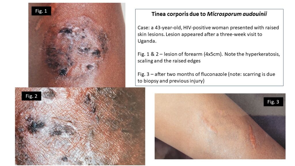
| NAMES Tinea corporis; ringworm; tinea gladiatorum (among wrestlers); tinea imbricata (AKA Tokelau) Reviewed by Leung et al (2020), including tinea incognito and tinea imbricata. |
| DISEASE – LESIONS: Non-symmetrical patches, often circular or oval, with a slightly raised red edge and scaling. Usually not itchy. The appearances may be quite different after application of local steroid ointment or other topical agents. Infections acquired from animals can be inflamed and pustular. – SITE: Usually on exposed areas of the extremities, but may be on the body or neck. |
| FUNGI Trichophyton rubrum; T. interdigitale, T. verrucosum; other Trichophyton spp. Microsporum canis; M. audouinii; Epidermophyton floccosum |
| GLOBAL BURDEN Worldwide and common, with a higher frequency in tropical and subtropical countries. Tinea imbricata is limited to Southeast Asia, India, southwest Polynesia, Melanesia, and Central America. |
| RISK FACTORS Transmitted by direct contact with other infected individuals or animals. M. canis infections acquired from dogs and other furry animals. Severe infections may occur in immunocompromised individuals |
| DIAGNOSIS Skin scraping with microscopy and fungal culture. |
| TREATMENT Reviewed by Sahoo & Mahajan (2016) Topical terbinafine cream over the lesions, oral terbinafine or itraconazole. |
| OUTLOOK Excellent. Recurrence is likely if continued contact with infected humans or animals. |


A lyrical awareness video on tinea corporis
Tinea capitis
| NAMES Tinea capitis; kerion; favus |
| DISEASE Tinea capitis appears as hair loss (alopecia) with little apparent inflammation. Well defined patches of hair loss start small and increase in size. In ‘black dot’ tinea capitis, hair breaks just above the scalp and diffuse swollen black dots appear. Clinical diagnosis requires the presence of broken hairs accompanied by scaling on the scalp, but can be difficult. Kerion is an inflammatory mass of hair, exudate, fungus and granulation tissue that can mimic a squamous cell carcinoma, and is more common in children. Favus is associated with red patches and scaling overlaid with disc or cup shaped yellow crusts (scutula) pierced by 1 or 2 hairs which do not break, with a distinctive, unpleasant odour. |
| FUNGI Microsporum audouinii; M. canis;, M. gypseum; several species of Trichophyton (T. interdigitale; verrucosum; T. tonsurans; T. soudanense; T. violaceum. Favus is caused by T. schoenleinii. |
| GLOBAL BURDEN Tinea capitis is worldwide in distribution, is more common in black adults and children with a global prevalence of 200 million cases. It is quite contagious and outbreaks may occur. In a recent US survey, tinea capitis was found in 6.6% children with ranges from 0% to 19.4%. It is more common in deprived areas. Favus is most common in remote areas of central and east Africa. Read a systematic review of global data by Rodríguez‐Cerdeira et al (2020) |
| RISK FACTORS Malnourished and deprived children are at greatest risk. Untreated infections may persist into adult life. |
| DIAGNOSIS Microscopy of hair roots and culture |
| TREATMENT Topical treatments are ineffective as they do not reach the inside of the infected hair shaft. The oral antifungals terbinafine, itraconazole and griseofulvin have similar efficacy, given for 2-6 weeks. M. canis infections are more difficult to treat and are refractory to terbinafine. |
| OUTLOOK Recurrence can occur if infected family cats or dogs are not tested and treated if infected. Hair loss (alopecia) is usually reversible, but may be permanent if the infection is longstanding, or with kerion or favus. |
| MORE INFORMATION Review on kerion by John et al (2016) LIFE lecture (parts 1 & 2) |


Tinea pedis
| NAMES Tinea pedis; athlete’s foot |
| DISEASE The most common infection is between the toes, especially between the 4-5th toes and 3-4th toes. Cracking with a painful fissure is common. Infection of the whole of the foot with a scaling eczema reaction and fissuring of the heel is also common (known as ‘moccasin type’). Occasionally an inflammatory reaction of the feet is seen. |
| FUNGI Trichophyton rubrum; T. interdigitale; rarely others |
| GLOBAL BURDEN Fungal infection of the skin, hair or nails affects ~25% of the world’s population (~1.5 billion) |
| RISK FACTORS Athlete’s foot affects anyone at any age, but is more common in those who have dampness between their toes related to sports, swimming, frequent bathing or hot climates without drying between the toe webs. Rates of up to 38% are seen among homeless persons (To et al, 2016). |
| DIAGNOSIS Microscopy and fungal culture |
| TREATMENT Terbinafine 1% cream or clotrimazole 1% cream. Oral itraconazole or terbinafine for moccasin type infection for 3 weeks |
| OUTLOOK Responds well to therapy but tends to recur. Lower leg cellulitis complicates athlete’s foot. |


Tinea cruris
| NAMES Tinea cruris; jock itch; genitocrural dermatophytosis |
| DISEASE LESIONS: Confluent, red, scaly rash. A common differential diagnosis is cutaneous candidiasis, in which satellite lesions are common. SITE: Generally covering the groin, scrotum and upper thighs. The central part of the rash may clear. The penile shaft is not affected. Intense pruritus (itching) is common. Localised scrotal infection is quite common and inconspicuous. There may be evidence of superficial fungal infection elsewhere on the body. |
| FUNGI Dermatophytes such as Trichophyton rubrum and Epidermophyton floccosum, but Candida albicans can also affect damp areas where intertrigo is a problem |
| GLOBAL BURDEN More common in men than women, and tends to occur between the ages of 18 and 60. Worldwide in distribution. Highly contagious; mini-outbreaks can occur e.g. in settings where towels are shared. |
| RISK FACTORS None especially, other than sharing of bathing facilities. |
| DIAGNOSIS Skin scraping with microscopy and fungal culture. |
| TREATMENT Local application of topical antifungal cream twice daily for 2-3 weeks is usually sufficient. If there is evidence of extensive infection then oral therapy with itraconazole or terbinafine is better. |
| OUTLOOK Excellent, although relapse occurs if other lesions on the body are not treated or treatment is not continued long enough. |


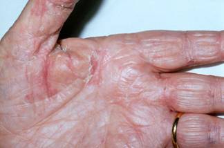
Tinea manuum
| NAMES Tinea manuum Read a more detailed guide at StatPearls or read a series of 18 Italian cases by Veraldi et al (2019) |
| DISEASE Usually unilateral, especially on the right hand. Always asymmetrical. Two distinct forms. The dyshidrotic (eczematoid) form presents with raised scaly or vesicular lesions and marked itching and burning. The hyperkeratotic form gradually gets worse with a gradually enlarging dry area of involvement leading to extensive involvement of most of the palm and fingers. Fissures and thickening of the skin making the hands rough is common. |
| FUNGI Trichophyton interdigitale;T. rubrum; Epidermophyton floccosum. Occasionally others. |
| GLOBAL BURDEN Worldwide, frequency unknown but uncommon among skin fungal infections. |
| RISK FACTORS None know; usually acquired from other infected individuals |
| DIAGNOSIS Skin scraping for culture and microscopy. |
| TREATMENT Topical azole therapy (econazole or clotrimazole) is usually successful, if applied for at least 10 days and possibly longer if extensive disease. Oral itraconazole (200 mg/d) or terbinafine (250 mg/d) are also highly effective, if given for 2-4 weeks. |
| OUTLOOK Excellent outlook. Fingernail involvement should also be sought and treated. |
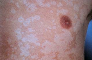
Pityriasis versicolor
| NAMES Tinea versicolor; pityriasis versicolor |
| DISEASE Superficial skin infection of the upper trunk, usually without an inflammatory component or scaling. Depigmentation, like rain drops, or excess pigmentation over the chest or upper back is typical, especially after sun exposure. |
| FUNGI Malassezia furfur (formerly Pityrosporum ovale) and other related species |
| GLOBAL BURDEN Common, especially in tropical and subtropical regions where up to 50% of the adult population may be affected. More obvious and perhaps more common in the summer months in temperate climates. Most common between ages of 20 and 40. |
| RISK FACTORS Possibly genetic predisposition as family members not living together are more frequently affected. |
| DIAGNOSIS Microscopy showing budding yeasts cells from the affected area. |
| TREATMENT There are several topical treatments available containing sulphide selenium, propylene glycol and azole antifungals. Extensive cases sometimes require oral azole treatment with ketoconazole or itraconazole. |
| OUTLOOK Rates of recurrence are very high. In some cases prophylactic itraconazole (200 mg one day per month for 6 months) has been used with a response of 88% at the end of the study. |