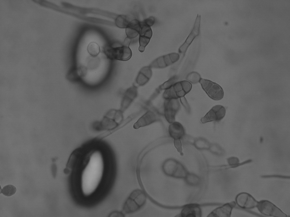
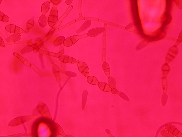
The majority of fungi form filamentous structure known as hyphae. These are multicellular structures with branching. Most of these hyphae extend in 3 dimensions through whatever they are growing in. Specialised hyphae are produced to allow vegetative (non-sexual) reproduction with spores or conidia. Some highly specialised reproductive or protective structures are also formed by some species, such as ascospores. There are probably millions of species in total. Filamentous fungi (also known as moulds) are found in most phylogenetic groups, but the vast majority of human pathogens are Ascomycetes.
- Jump to: Aspergillus, Fusarium, Lichtheimia, Rhizopus, Trichophyton, Alternaria, Apophysomyces, Cladosphialophora, Fonsecaea

Factsheets
Alternaria
| NAMES Alternaria alternata |
| NATURAL HABITAT Found in soil, air and it has also been described on normal human and animal skin and conjunctiva. |
| GEOGRAPHY Worldwide |
| FREQUENCY It is frequently associated with allergic respiratory disease. It is an infrequent case of localised and disseminated infections. |
| DISEASES – Allergic respiratory disease, including thunderstorm asthma and SAFS (watch an interview with a patient with allergic bronchopulmonary mycosis) – Allergic fungal rhinosinusitis – Opportunistic infections in immunocompromised hosts, including in skin, paranasal sinuses and lungs. – Keratitis, peritonitis, osteomyelitis, sinusitis, endophthalmitis, and cutaneous and subcutaneous infections associated with a previous trauma. |
| CULTURE Fast-growing colonies, grey to black and powdery to floccose. Microscopically, conidiophores arise from septate hyphae. Multicellular conidia are produced sympodially in chains (usually composed of more than 5 conidia). Conidia are obclavate, pyriform to ovoid or ellipsoidal up to 50 mm long and 3-12 mm wide. Brown pigmented with smooth to verrucose surface. Biosafety level 2 |
| ANTIFUNGAL RESISTANCE This species is resistant to fluconazole. Resistance to itraconazole and voriconazole have been described and some strains have shown in vitro resistance to amphotericin B. |
| INDUSTRIAL USES Biocontrol of some weed plants. |

Apophysomyces
| NAMES Apophysomyces species (A. elegans, A. ossiformis, A. trapeziformis, A. variabilis) are clinically identical and difficult to distinguish by traditional diagnostics, so they are included together here. |
| NATURAL HABITAT Isolated in soil and decaying plant debris. |
| GEOGRAPHY Worldwide, but more frequent in tropical and subtropical areas |
| FREQUENCY Unlike other Zygomycetes, frequency of infection is more common with immunocompetent hosts. The real frequency is unknown but it is far more common in tropical and subtropical areas. A. trapeziformis has recently been identified as the cause of mucormycosis in 13 survivors of the Joplin tornado in Missouri, USA (read the story at Medscape). |
| DISEASES Most commonly cutaneous and subcutaneous infections following the traumatic implantation of the spores. Sinus infections have also been described. |
| CULTURE Great expertise is required to differentiate the three species of the complex. Experts can distinguish them by the morphology of their sporangiophores and sporangiospores. It is advisable to confirm identify by means of DNA sequencing the ITS fragment. Fast growing; pale white turning brownish-grey with age. Microscopically, pyriform sporangia, apophyses funnel or bell-shaped. Sporangiospores are different depending on the species being bone-shaped, trapezoid-shaped or variable-shaped. Biosafety level 2 |
| ANTIFUNGAL RESISTANCE All isolates are intrinsically resistant to fluconazole, ketoconazole, voriconazole and the echinocandins. Usually susceptible to amphotericin B and posaconazole. Variably susceptible to itraconazole. |
| INDUSTRIAL USES None |

Cladosphialophora
| NAMES Cladosphialophora spp. Includes C. carrionii, Cladosphialophora bantiana, and others. |
| NATURAL HABITAT Soil and rotten plant material |
| GEOGRAPHY Worldwide distribution, especially semi-arid regions |
| FREQUENCY Unknown |
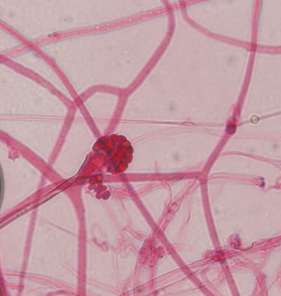
| DISEASES Cladosphialophora spp. are causative agents of phaeohyphomycosis, chromoblastomycosis, and mycetoma. Species of this genera have been isolated from cutaneous, subcutaneous and disseminated infections. C. bantiana causes cerebral phaeohyphomycosis in immunocompetent patients. |
| CULTURE Colonies restricted powdery to woolly, olivaceous green to black. Conidiophores are mostly absent or not well developed; unicellular conidia in chains without pigmented scares. Biosafety level 2: C. carrionii Biosafety level 3: C. bantiana |
| ANTIFUNGAL RESISTANCE Intrinsically resistant to fluconazole. High MICs to echinocandins and some strains show high MICs to amphotericin B. |
| INDUSTRIAL USES None |

Fonsecaea
| NAMES Fonsecaea pedrosoi complex which includes F. monophora and the previously named species F. compacta, now incorporated in F. pedrosoi complex. [ Teleomorph = Hormodendrum pedrosoi ] |
| NATURAL HABITAT Rotten wood and soil |
| GEOGRAPHY Worldwide distribution, especially in humid areas. |
| FREQUENCY Unknown |
| DISEASES It is the main agent of human chromoblastomycosis. It has also been isolated from cases of keratitis, paranasal sinusitis and brain abscess. |
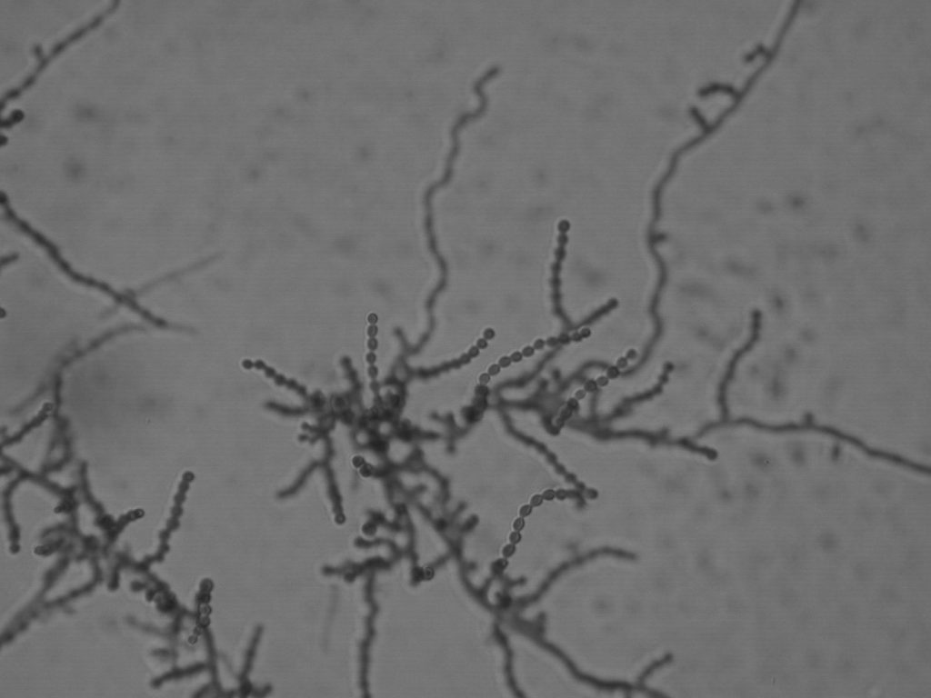
| CULTURE Colonies are slow growing, lanose to velvety, olivaceous to black. Microscopically it is characterized by dark brown hyphae and suberect conidiophores loosely branched. Phialides with funnel-shaped collarettes and/or simpodial growth might be present. All strains grow at 37C but not at 40C. Biosafety level 2 |
| ANTIFUNGAL RESISTANCE Some strains show elevated MICs to AMB. Echinocandins have none or low activity against this species. |
| INDUSTRIAL USES None |
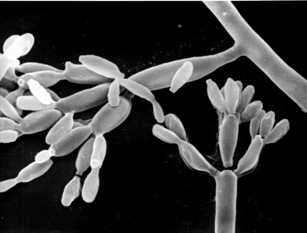
Scanning electromicrograph image of F. pedrosoi complex. 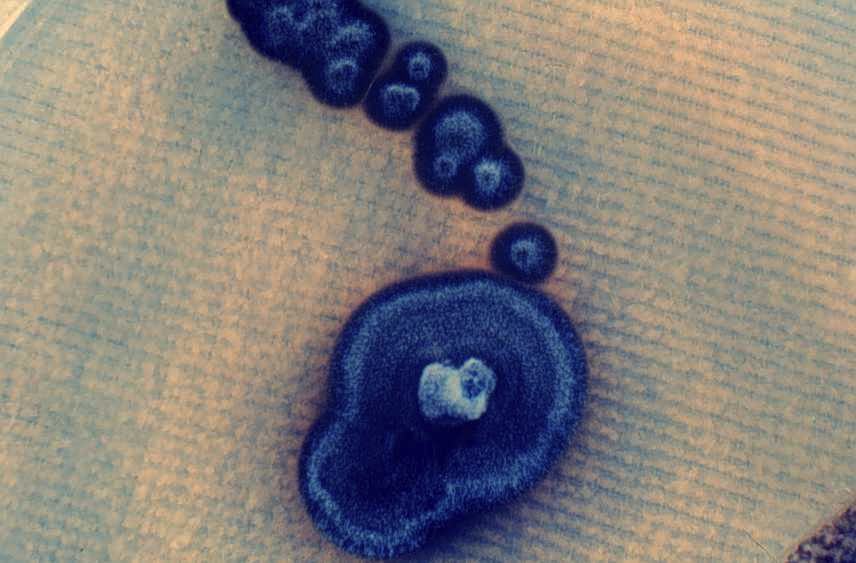
High power image of a typical image of highly characteristic ‘muriform’ cells seen histologically during infection. Muriform bodies have a brown (melanin) surface layer and are seen within a chronic inflammatory infiltrate with granulomas and micro abscesses. 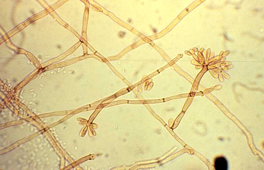
F. pedrosoi complex.

