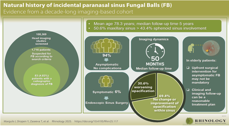
A remarkable paper from Itai Margulis and colleagues in Petah Tikva, Israel, identified an incidental fungus ball in the sinuses of 83 patients among 108,360 head CT scans (0.075%) over 12 years. The maxillary sinus was involved in 42 (50.6%), and the sphenoid in 36 (43.4%) patients and 81 (97.6%) were unilateral. Most of the patients were older people, with a mean age of 78 years, although some were in their 30’s, and 42% had diabetes.
Even more remarkable, only 5 (6%) of patients underwent endoscopic surgery and none, open surgery. Follow up scans were done in 49 patients at least 6 months later, and some as long as 11 years later (median 4 years). In 29 (59.2%) there was no change and in 5 (10.2%) a decreased opacity within the sinus. Increased opacity within the sinus was seen in 13 (26.5%) and increased opacity outside the sinus in 2 (4.1%).
The usual practice if a fungal ball is detected in a sinus is to remove it, if there are any symptoms referable to it. This paper casts doubt on this course of action, at least in older patients who are asymptomatic.
The first record of any human case of (probable) aspergillosis relates to the removal of a progressive fungal ball from the maxillary sinus in a soldier in Paris [https://www.aspergillus.org.uk/historical-papers/]. Brass sutures were used, along with hot cautery. The operation was a success.