Most imaging of fungal infection is done with plain chest radiology or CT scanning.
An increasing number of imaging investigations use MR, especially for the brain. Sometimes ultrasound is useful especially for cardiac echocardiography (endocarditis) and sometime direct visualization of fungi is possible down a bronchoscope (tracheobronchitis) or endoscope (oesphageal candidiasis or acute invasive fungal rhinosinusitis). Imaging is important for initial diagnosis and for following progress of infection over time. Simple enlargement of abnormal areas does not necessarily mean clinical failure, as enlargement can represent immune reconstitution, in some patients.
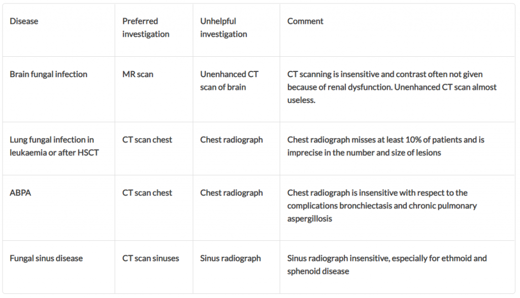
Some radiology investigations are insensitive or unhelpful. These are listed below:
Setting the correct parameters for CT and MR scanning requires radiological expertise. Likewise interpretation is critical and can be difficult. Radiological imaging cannot make a precise mycological diagnosis but usually contributes greatly to a full understanding of the extent of disease and its treatment. Mistaken radiological diagnosis does occasionally occur, primarily because a wide differential diagnosis is not considered initially, and some fungal infections are rare.