Often the most direct and conclusive way of identifying the fungus causing an infection is to grow it from a patient sample, such as blood, CSF, pus, urine, tissue, respiratory samples (sputum, BAL), pleural, pericardial or peritoneal fluid, skin scraping, hair, nail clippings, oral or vaginal samples.
Sample processing may involve centrifugation or softening/liquidisation to allow the sample to be spread onto agar. A number of laboratory manuals and specific describe the methods for doing this although remarkably few comparative studies comparing one method with another have been done.
Medium selection
FUNGAL MEDIA: Yield is usually improved by direct culture on specialist fungal media, for example culture of Aspergillus spp. on bacterial media is ~30% less effective than on fungal media (Horvath & Dummer, 1996), and some fungi (e.g. Histoplasma, Mucorales and Coccidioides spp.) will not grow on bacterial media. General purpose media that are commonly used for fungal culture are Sabouraud dextrose, malt extract, and less commonly brain heart infusion medium.
BLOOD CULTURE SYSTEMS: Numerous blood culture systems have been used, including ‘standard’ bacterial cultures and more specialised systems such as the lysis centrifugation system, the SEPTI-CHEK systems, and special bottles within an automated system (sometimes of Mycobacterium and fungi). A small number of comparisons have been done of these systems, and the choice matters a great deal more when the range of potential infecting fungi includes Cryptococcus neoformans, Histoplasma capsulatum and Penicillium marneffei. In current automated blood culture systems, Candida glabrata grows more slowly and much more often in the anaerobic bottle, so both aerobic and anaerobic bottles should be filled with blood. The yield from blood culture is higher if patients are not taking antifungal agents (Kami et al, 2002) and if at least 20 ml of blood is cultured.
SELECTIVE MEDIA: specialist media can be used to distinguish between similar-looking colonies:
- Bird seed agar to identify Cryptococcus spp. in respiratory cultures from HIV patients
- Chromogenic agar (e.g. CHROMagar) for the direct, partial identification of Candida spp.
CYCLOHEXIMIDE can be added to reduce contamination by environmental fungae, but this reduces the yield of many opportunistic fungi (e.g. Aspergillus spp., Cryptococcus neoformans and Mucorales isolates), so one agar plate not containing it should also be used in parallel.
CHLORAMPHENICOL can be used to prevent bacterial contamination, but prevents the growth of Actinomyces, which others grows well on Sabouraud dextrose agar.
INCUBATION: Standard conditions are incubation at 30ºC in a humidified environment for 21 days. Inspect daily for at least a week, and at least 3 times weekly thereafter. Some fungi are very slow to grow (e.g. Histoplasma spp.) and may need longer incubation times. Incubate respiratory cultures at 42ºC to select for Aspergillus spp. as this prevents the growth of Candida spp.
A research paper published by Zhu et al in 2021 showed that molecular detection, rather than length of incubation, had most impact on the diagnosis of fungal infections. In their study 96% of positive cultures were identified within seven days.
Biosafety

Plates should not be opened outside a secure environment if there is any possibility of potentially airborne pathogens such as Coccidioides spp., Histoplasma spp., Blastomyces dermatitidis, Penicillium marneffei. Laboratory transmission of infection has occurred with severe consequences. Measures for preventing and dealing with accidental laboratory exposure to Coccidioides (a biosafety level 3 organism) are described in Stevens et al, 2009.
Identification
Some fungi can be identified based on their culture characteristics while others will need additional tests such as MALDI-ToF, molecular tests or
Automated systems such as the VITEK2 (bioMerieux, includes yeast ID cards) are also an option for larger laboratories and may be able to incorporate antifungal susceptibility testing.
IDENTIFYING YEASTS: Yeasts can be further identified by their growth pattern on specialised media such as cornmeal agar and with biochemistry testing. Some presumptive identification of yeasts can be made based on colour and colony morphology on chromogenic media. Some rapid testing systems are available which are more or less comprehensive and reliable. Published comparisons of these methodologies are available.
IDENTIFYING FILAMENTOUS FUNGI: These are harder to identify. Without spores or other sterile structures, the only real distinction that can be made is whether the hyphae are septate or not (i.e. are likely be Mucorales, Basidiobolus ranarum or Conidiobolus spp). Media that can assist with sporulation include potato dextrose agar. Once the colony has sporulated, identification by microscopy using phenol cotton blue or another simple means of highlighting the distinctive structures of the fungus allows an experienced mycologist to identify to at least genus level in the vast majority of cases. If the colony will not sporulate, only molecular identification is possible. Species identification of filamentous fungi can be very difficult or impossible without additional data such as colour and morphology of additional media, differential temperature growth rates and molecular information. Many cryptic (i.e. identical appearance) species have been described in many genera.
Interpreting culture results
DIAGNOSIS: culture of most fungi from a human sample makes the diagnosis as external contamination is rare. So a positive culture of Trichophyton spp., Microsporum spp. Cryptococcus neoformans, Coccidioides spp., Histoplasma spp., Blastomyces dermatitidis, Penicillium marneffei represent disease.
CONTAMINANTS: The main exceptions are Aspergillus spp. and Penicillium spp., and occasionally other pathogenic fungi such as the bread mould Rhizopus, are plate contaminants or found in specimens but not causing disease. In addition, Candida albicans may be cultured from the mouth or urine or sputum and not be pathogenic at the time of culture.
For further information see Borman & Johnson (2014)
| Aspergillus | Candida | Penicillium | |
| Blood | Rare, may be significant or laboratory contaminant | Pathogenic and requires treatment (rare exceptions related to skin colonisation) | Not significant, a laboratory contaminant |
| Urine | Rare, possibly significant | Usually not significant. In immunocompromised or intensive care may signal disseminated candidiasis | |
| CSF | SIGNIFICANT | SIGNIFICANT | Probably laboratory contamination |
| Pus, or sterile site aspiration | SIGNIFICANT | SIGNIFICANT | Rare, probably significant, could be laboratory contaminant |
| Sputum, or other respiratory samples | Often significant in allergic, chronic or invasive disease, especially if A. fumigatus | Rarely significant | Not significant. Possibly significant in allergy. |
| Peritoneal discharge after surgery | Rare, probably laboratory contaminant | Often significant, for Candida peritonitis | Rare, probably laboratory contaminant |
| CAPD fluid | Rare, possibly significant | SIGNIFICANT AND SERIOUS | Rare, probably laboratory contaminant |
| Venous catheter | Rare, may be significant or laboratory contaminant | Probably significant – bloodstream infection likely, may be silent | Not significant, a laboratory contaminant |
| Tissue | SIGNIFICANT if confirmed by histology | SIGNIFICANT if confirmed by histology | SIGNIFICANT if confirmed by histology |
| Cornea scraping | Probably significant | Probably significant | Rare, probably laboratory contaminant |
| Ear swab | Probably significant if A. niger; consider invasive otitis if A. fumigatus | Probably significant | Laboratory or skin contaminant |
| Nails | Probably significant, if toenails | Probably significant, if nail fold or clinical appearance is that of superficial white onychomycosis | Laboratory or skin contaminant |
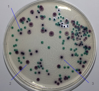
ChromAgar
Chromogenic agar is available for Candida and Malassezia, which can differentiate the major species with very high specificity. Material can be directly used for MALDI-ToF without further subculturing.
Chromogenic agar suppliers:
www.sigmaaldrich.com/GB/en/product/sial/94382
www.chromagar.com/en/product/chromagar-candida/
www.oxoid.com/UK/blue/prod_detail/prod_detail.asp?pr=CM1002&c=UK&lang=EN
www.biomerieux-diagnostics.com/culture-media-fungal-infections
www.neogen.com/en-gb/categories/microbiology/harlequin-candida-chromogenic-agar/
www.tmmedia.in/product/chromogenic-candida-agar-chromogenic-candida-differential-agar/
Candida albicans → green-blue
Candida auris → light blue with blue halo
Candida tropicalis → metallic blue with pink halo
Candida krusei → pink and fuzzy
ChromAgar: 1=Candida albicans; 2= Candida krusei; 3=Candida tropicalis 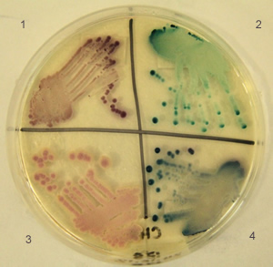
ChromAgar :1=C. glabrata; 2= C. albicans 3=C. krusei; 4=C. tropicalis